No difference in rate of overall positive margins for all lesions; size, morphology impact positivity
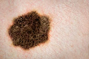
FRIDAY, April 17, 2015 (HealthDay News) — For clinically atypical pigmented lesions, lesion size and morphology should be considered before deciding on shave or punch biopsy, according to a research letter published online April 9 in the British Journal of Dermatology.
R. Cheng, from the Duke University School of Medicine in Durham, N.C., and colleagues conducted a retrospective chart review to examine the rate of positive margins for punch and shave biopsies of clinically atypical pigmented lesions. The authors included data from 607 consecutive biopsies, mainly dysplastic nevi.
The researchers observed no difference in the rate of positive margins between shave and punch biopsies when including all lesions (21.2 versus 22.9 percent; P = 0.688). For larger lesions (5 to 8 mm in diameter), the percentage of overall positive margins was lower for shave than punch biopsies (P = 0.032). For smaller lesions (0 to 4 mm in diameter) and larger lesions (5 to 8 mm in diameter), shave biopsies had a significantly lower rate of lateral positive margins (P = 0.008 and P = 0.002, respectively). Shave biopsies resulted in significantly lower rates of positive lateral margins for all macular lesions (P = 0.036), and for larger macular lesions (P = 0.034). For all lesions, the rate of positive deep margins was significantly lower for punch versus shave biopsies (P < 0.0001).
“Lesion size and morphology should be considered when clinically atypical pigmented lesions are biopsied,” the authors write.
The study was partially funded by Research Electronic Data Capture tools.
Copyright © 2015 HealthDay. All rights reserved.


