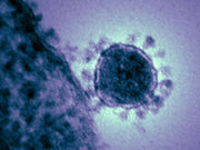
Main finding in lungs was diffuse alveolar damage; evidence of chronic disease in other organs
FRIDAY, Feb. 5, 2016 (HealthDay News) — The main histopathologic findings of Middle East respiratory syndrome coronavirus (MERS-CoV) infection are diffuse alveolar damage in the lungs and evidence of chronic disease in other organs, according to research published online Feb. 5 in The American Journal of Pathology.
Dianna L. Ng., from the U.S. Centers for Disease Control and Prevention in Atlanta, and colleagues describe the histopathologic, immunohistochemical, and ultrastructural findings from the first autopsy performed on a fatal case of MERS-CoV. The fatality was related to a hospital outbreak in the United Arab Emirates in April 2014.
The researchers note that diffuse alveolar damage was the main histopathologic finding in the lungs. In other organs there was evidence of chronic disease, including severe peripheral vascular disease, patchy cardiac fibrosis, and hepatic steatosis. Pneumocytes and epithelial syncytial cells were identified as important targets of MERS-CoV antigen in double staining immunoassays that used anti-MERS-CoV antibodies paired with immunohistochemistry for cytokeratin and surfactant. Colocalization in scattered pneumocytes and syncytial cells was seen in double immunostaining with dipeptidyl peptidase 4. There was no evidence of extrapulmonary MERS-CoV antigens, including in the kidney.
“These results provide critical insights into the pathogenesis of MERS-CoV in humans,” the authors write.
Copyright © 2016 HealthDay. All rights reserved.


