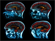
Increased iron concentration seen in deep gray matter, neocortical regions versus healthy controls
TUESDAY, June 30, 2020 (HealthDay News) — Patients with Alzheimer disease (AD) have increased iron concentration in the deep gray matter and neocortical regions compared with healthy controls (HC), according to a study published online June 30 in Radiology.
Anna Damulina, M.D., from the Medical University of Graz in Austria, and colleagues examined baseline and change in brain iron levels using magnetic resonance imaging (MRI) with R2* relaxation rate mapping in 100 individuals with AD, recruited between 2010 and 2016, and 100 age-matched HC, selected from 2010 to 2014. All participants underwent 3-Tesla MRI, including R2* mapping. At a mean follow-up of 17 months, 56 of 100 participants with AD underwent neuropsychological testing and brain MRI.
The researchers found that compared with the HC group, the AD group had higher median R2* levels in the basal ganglia and total neocortex, and regionally in the occipital and temporal lobes. In participants with AD, there was an association between R2* values in the temporal lobe and longitudinal changes in Consortium to Establish a Registry for Alzheimer’s Disease total score, independent of longitudinal changes in brain volume.
“Our study provides support for the hypothesis of impaired iron homeostasis in Alzheimer’s disease and indicates that the use of iron chelators in clinical trials might be a promising treatment target,” a coauthor said in a statement. “MRI-based iron mapping could be used as a biomarker for Alzheimer’s disease prediction and as a tool to monitor treatment response in therapeutic studies.”
One author disclosed financial ties to the pharmaceutical industry.
Copyright © 2020 HealthDay. All rights reserved.


